
The large high-resolution flat panel detector has an input resolution equivalent to 14 megapixels, which provides a large field-of-view and high resolution.
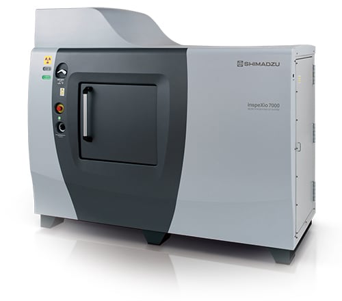
Microfocus X-Ray CT System
The inspeXio 7000 is a high-performance microfocus X-ray CT system equipped with a Shimadzu microfocus X-ray generator and a large high-resolution flat panel detector.
The large detection area, input resolution equivalent to 14 megapixels, and an enhanced high-output microfocus X-ray generator enable CT images with a large field-of-view, high resolution, and high contrast. In addition, the improved HPCinspeXio high-performance computing system processes images faster.
These developments make the inspeXio 7000 system applicable for researching, developing, or inspecting a wide variety of samples, from composite materials, such as glass fiber reinforced plastic (GFRP) and continuous fiber reinforced thermoplastic laminate (CFRTP) materials to large aluminum die cast parts.
The Analytical Intelligence logo and CORE Boost are trademarks of Shimadzu Corporation or its affiliated companies in Japan and/or other countries.
VGSTUDIO MAX and VGSTUDIO are trademarks of Volume Graphics GmbH.
POLYGONALmeister is a trademark of UEL Corporation.
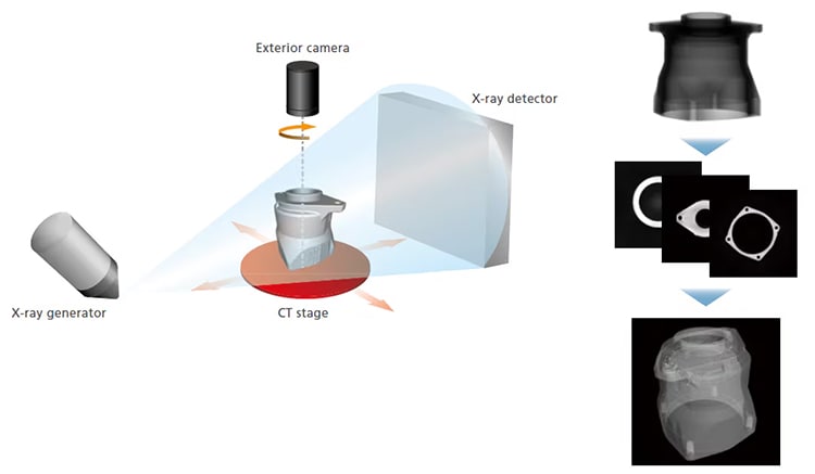
The inspection target (sample) is placed between the X-ray generator and detector, as shown below. Then, the sample is rotated 360 degrees to collect X-ray fluoroscopic data from various angles in order to calculate cross-sectional images.
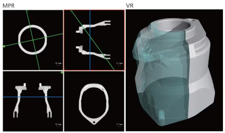
Displays any cross section desired
Multi Planar Reconstruction (MPR) stacks multiple CT images in a virtual space to display four images—a CT image, mutually longitudinal section images, and a user-selected section image orthogonal to one of the longitudinal section images.
Volume rendering (VR) stacks multiple CT images in a virtual space to display a 3D image. Separate 3D image processing software is required for VR display.