ASMS (American Society for Mass Spectrometry) 2025
June 1-5
Baltimore Convention Center
Baltimore, MD
The majority of proteins synthesized in biological organisms undergo glycosylation. Glycosylation is modification with glycans of high structural heterogeneity composed of multiple monosaccharides, such as glucose and galactose, which are bonded together. These glycans classified into N-linked types and O-linked types. These types of glycan are known to play an important role in regulating protein function, and obtaining information on protein glycosylation is essential to the development of biopharmaceuticals.
One piece of this information related to glycosylation is where the glycan is bonded to the protein. Almost all N-linked glycans have an amino acid consensus sequence of -NXS/T-, which is used to discover potential N-linked glycan binding sites. N-linked glycan binding sites can be determined by digesting a glycoprotein with a protease, collecting glycopeptides from the digested material using lectin that binds specifically to glycans, cleaving the glycans from the peptides using the N-glycosidase (PNGase F) in the presence of H218O, labeling the N-linked glycan binding sites with a stable isotope, and then performing analysis.1) N-linked glycan binding sites can also be determined by MSn analysis using product ions that are specific to N-linked glycopeptides.2) The group of glycans called O-linked glycans are known to bond to a serine or threonine amino acid on the protein. Though often little information is available on the amino acid sequence around this O-linked glycan binding area, there is no enzyme suitable for cleaving the bond as with N-linked glycans, and also no specific product ions have yet been discovered, which makes determination of the binding site of O-linked glycans difficult.
See how Shimadzu provides a variety of solutions for your glycan characterization needs.
The Raptor Polar X column was chosen in this study due to its unique stationary phase which balances ion exchange and hydrophilic interaction liquid chromatography (HILIC) to retain and separate polar analytes. This balanced retention for polar compounds should provide a powerful separation of glycans. The released glycan data was processed using Detached Glycan (N-Linked) Workflow from Protein Metrics. This workflow can be combined with previously published workflows for complete mAb characterization.
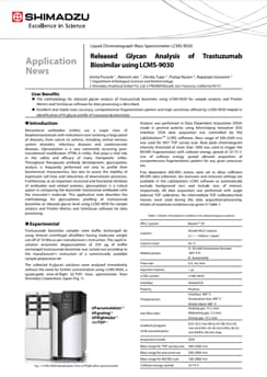
Monoclonal antibodies (mAbs) are a major class of biopharmaceuticals with indications now covering a large panel of diseases, from cancer to asthma, including central nervous system disorders, infectious diseases and cardiovascular diseases. Glycosylation is a very commonly occurring posttranslational modification (PTM) in mAbs, that plays a vital role in the safety and efficacy of many therapeutic mAbs. Throughout therapeutic antibody development, glycosylation analysis is frequently performed not only to profile their biochemical characteristics, but also to assess the stability of expression cell lines and robustness of downstream processes. Furthermore, as an important structural and functional attribute of antibodies and related proteins, glycosylation is a critical aspect in comparing the biosimilar monoclonal antibodies with the innovator’s molecule. This application note describes the methodology for glycosylation profiling of trastuzumab biosimilar at released glycan level using LCMS-9030 for sample analysis and Protein Metrics and SimGlycan software for data processing.
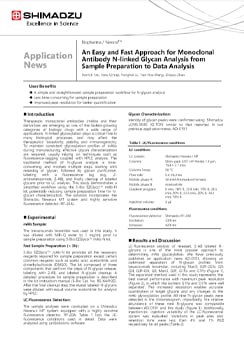
Therapeutic monoclonal antibodies (mAbs) and their derivatives are emerging as one of the fastest-growing categories of biologic drugs with a wide range of applications. N-linked glycosylation plays a critical role in many biological processes, and may affect the therapeutics’ bioactivity, stability and immunogenicity. To maintain consistent glycosylation profiles of mAbs during manufacturing, effective glycan characterization are required, usually relying on techniques such as fluorescence-tagging coupled with HPLC analysis. The traditional method of N-glycan analysis is time consuming, and involves multiple steps, starting with releasing of glycan, followed by glycan purification, labeling with a fluorescence tag (e.g., 2- aminobenzamide, 2-AB), and finally cleanup of labeled glycans prior to LC analysis. This study demonstrates a simplified workflow using the S-Bio EZGlycoTM mAb-N kit, potentially reducing sample preparation time for Nglycan characterization. The solution incorporates the Shimadzu Nexera-i MT system and highly sensitive fluorescence detector (RF-20A).
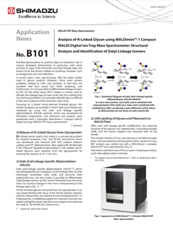
N-linked glycosylation to proteins plays an important role in various biological phenomena. In particular, sialic acids existing at sugar chain terminals and their linkage types are known to be key factors related to numerous diseases, such as antigenicity and viral infections. In recent years, mass spectroscopy (MS) has been widely used in glycan analysis. However, there were various problems related to sialic acid residues, in that they are unstable and thus easily lost while analysing, and furthermore, it is not possible to differentiate linkage isomers by MS. On the other hand, HPLC analysis is mainly used to identify the linkage type of sialic acids, but this method also has technical problems, for example, identification is difficult in the case of glycans with numerous sialic acids. Focusing on a blood serum-derived N-linked glycan, this article introduces an example in which sialic acid residue was stabilized by using the sialic acid linkage specific alkylamidation (SALSA) method, which was developed by Shimadzu Corporation, and detection and analysis were performed with a Shimadzu MALDImini-1 compact MALDI digital ion trap (MALDI-DIT) mass spectrometer.
Many protein-based biopharmaceutical products, typified by antibody drugs, are synthesized in cultured cells derived from eukaryotes such as CHO (Chinese hamster ovary) cells. For this reason, there are inevitably many post-translational modifications to the biosynthesized proteins. Among these, modifications of glycans have gained attention as items for evaluating the quality of biopharmaceuticals since they are associated with the adjustment of protein functions, as well as with the unwanted development of antigenicity depending on their structure. However, there are various technical challenges in evaluating glycans. With O-linked glycans (O-glycan), it is particularly difficult to comprehensively release glycans from protein using enzymes, leading to the use of mainly the following two chemical methods: hydrazinolysis and β-elimination. However, these methods have issues that need to be improved. Hydrazinolysis requires great care since an explosive reagent is handled and therefore is not easy to implement. With the β-elimination method, a peeling reaction where glycans gradually decompose due to a continuous elimination reaction occurs. Conventionally, in analysis of O-glycans using βelimination, glycans are released so as not to cause a continuous elimination reaction by using the reductive β-elimination method, which involves simultaneous releasing of glycans under alkaline conditions and reducing the root portion of the glycan with a reducing agent. However, this method completely reduces the root portion of the glycan and therefore does not allow labeling such as with fluorescent reagents after releasing glycans, thus limiting the available analysis methods. Also, in analysis by mass spectrometry of glycans obtained in this way, the high sensitivity analysis is not possible because the ionization efficiency of the glycan itself is not high. To address this, a non-reductive β-elimination/fluorescent labeling method is being examined as a method to bind a fluorescent labeling reagent such as 2-AB or PA by not reducing the root of the glycan, but this has not succeeded in significantly suppressing the continuous elimination reaction. Even so, in academic researches where O-glycan was the object of analysis, the existence of by-products due to this peeling reaction has not been problematic enough to hinder researches. However, glycans have to be evaluated for quality control in drugs which are to be administered to the human body, such as biopharmaceuticals, and the question of how to handle the existence of by-products during this evaluation is a major issue. In this article, we report the results of studying a method for releasing O-glycans chemically in which the peeling reaction is suppressed, based on a PMP labeling method.
Many protein-based biopharmaceutical products are synthesized in cultured cells derived from eukaryotes. For this reason, the synthesized proteins are mainly glycoproteins which comprise proteins with glycans linked to them. The glycans in these glycoproteins are broadly divided into N-linked glycans (N-glycans) and O-linked glycans (O-glycans), each having diverse and complex branching structures. The structure of the glycan is known to affect the function and stability of the glycoprotein. Therefore, if the structure of the glycan in a synthesized glycoprotein changes, due to changes in the culture environment for example, there may be unexpected changes in the function and stability of the glycoprotein itself. This possibility can lead to serious problems in the development and manufacture of biopharmaceutical products, and therefore monitoring whether the glycan structure has changed or not is a primary element in managing quality. It is important to correctly analyze and evaluate changes in glycan structure, but there are a variety of pretreatment methods for glycan analysis and they are not standardized, so the result of analysis of the same glycoprotein may differ if different pretreatment methods are used. This article introduces the results of comparing some of the pretreatment methods widely used in N-glycan analysis and investigating how they affect the analysis results.
Protein glycosylation has been shown to be related to protein activity. It is therefore of great interest to study glycan structures of glycoproteins in immunology and cellular biology. On top of that, it is also essential to analyze glycans in recombinant glycoprotein drugs to ensure consistent glycosylation profile. Glycan can be linked to protein either via asparagine (N-glycan) or via serine/threonine (O-glycan). Previously, we have published an application news on N-glycan analysis. 1 Typical analysis of glycan involves cleavage of the glycan from the protein and subsequently derivatization with fluorescent labels (e.g., 2-aminobenzamide, 2-AB) followed by analysis using HPLC. Unlike N-glycans which have well-established glycan releasing strategy, the Oglycans lack practical method for releasing of intact Oglycans. O-glycan releasing method such as hydrazinolysis often results in large amount of side reaction peeling products and requires long preparation time. This study demonstrates a simplified workflow using the S-Bio EZGlycoTM O-Glycan Prep kit, which exhibits low peeling and a reduced sample preparation time for O-glycan characterization. The solution incorporates the Shimadzu Nexera-i MT system and highly sensitive fluorescence detector (RF-20A).
ASMS (American Society for Mass Spectrometry) 2025
June 1-5
Baltimore Convention Center
Baltimore, MD
Shimadzu Scientific Instruments Opens Boston Location of Its R&D Center Focus will be on promoting customer-oriented development to expand business in the pharmaceutical field
Shimadzu Scientific Instruments, Inc. (SSI, Columbia, Maryland, USA), a Shimadzu Group company, has opened a satellite lab in Boston, Massachusetts to be the base of its collaborative research and development activities on the East Coast. Established to conduct research and development more closely linked to customers, SSI's R&D Center consists of three bases, with the main facilities at its Maryland headquarters, a West Coast location, and this new space in Boston near the city center. The Boston lab was set up by partnering with Labshares, a shared laboratory service provider for life science companies.