MALDI Theory and Basics
| Navigation |
|---|
| I. MALDI Theory |
| II. TOF MS Analysis |
| III. TOF/TOF CID Analysis |
| IV. MALDI mini – Digital Ion Trap |
| V. Atmospheric MALDI |
| MALDI Products and Solutions |
I. MALDI Theory
Mass spectrometry is an analytical technique used to measure the mass-to-charge ratio of ions from an analyte of interest. Different techniques are used depending on the characteristics of the analyte of interest to aid in detection. Matrix assisted laser desorption ionization (MALDI) is one technique and has become a powerful tool for the analysis of peptides and proteins since its introduction by Karas and Tanaka1, 2, 3. This technique combines an energy absorbing compound (matrix) with the analyte of interest. When the latter two compounds are mixed, it facilitates the desorption process for ionization and detection.
MALDI is a soft ionization technique that utilizes an energy absorbing matrix to aid in analyte detection. The analyte of interest is mixed with a matrix with a ratio that can vary from 1:100 to 1:50,000. The excess of matrices allows for an ease of energy transfer upon excitation from a laser4, 5, 6. Traditional MALDI matrices absorb in the ultra-violet (UV) range and are optimized for laser source laser emission wavelengths (nitrogen laser light, 337 nm, Nd:YAG laser, 355 nm)7 . Existing matrices have been chosen for their ability to absorb energy at these specific wavelengths and new matrices are optimized to absorb efficiently in this range. As such, the more commonly used matrices have functionalized aromatic compounds that can transfer or absorb a proton efficiently. Laser photon energy absorption generates heat in the matrix/analyte mixture, causing the matrix to vaporize along with the analyte of interest. Figure 1 shows a depiction of the matrix and analyte molecules being vaporized into a gas plume after excitation with a laser beam.
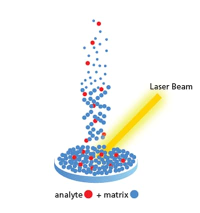
Figure 1. Sample irradiation with laser source, causing both matrix and analyte to vaporize.
After the analyte of interest has been vaporized, there are multiple ionization pathways possible for the analyte to proceed towards the detector8. The two most discussed models are the desorption of preformed ions or “Lucky Survivor” model9 and gas phase protonation model10 (Scheme 1). Desorption of preformed ions is common due to MALDI sample preparation. Trifluoroacetic acid is added at a low concentration to drop the matrix solution to a pH of 2-3, thus causing the sample to contain charged species when combined for analysis. When these charged ions are vaporized, they can collide with a variety of ions, neutral ions, including anions or cations. These different collision pathways can result in analyte neutralization or proton transfer of the analyte of interest. The second mode of ionization, gas phase protonation relies on the collision of a neutral analyte ion with a protonated or deprotonated matrix ion. Regardless of the ionization mode, the same mass is detected. The type of ion is highly dependent on the pH of the analyte solution. Acidic solutions produce more preformed ions for detection while basic solutions see more gas phase protonation11.

Scheme 1. Protonation mode for gas phase protonation and precharged analyte modes. Where ‘A’ is the analyte of interest, ‘H’ is a proton and ‘X’ is an anion.
II. TOF MS Analysis
MALDI analysis is often paired with a Time of Flight (TOF) mass analyzer. This allows for separation of ions based on the time (t) it takes to reach a detector at the end of a fixed distance (D) which corresponds to the length of the field free drift region. When ions are formed, they are accelerated using a high voltage and brought to the same kinetic energy. This kinetic energy is proportional to the accelerating voltage V as shown in equation (1)12
½ m v2 = eV (1)
and v = D / t (2)
Where, (m) is the ion molecular weight, (v) is the speed of the accelerated ion, (e) is the ion charge, and (V) is the value of the accelerating voltage. By combining equations (1) and (2) we can deduce equation (3) that defines the time of an ion flight through the drift tube.
t = (m / 2eV) ½ D (3)
By measuring the time it takes an ion to travel in the flight tube and reach the detector, we can deduce the value of its mass to charge ratio. Time of flight analyzers have been coupled with MALDI ionization sources to aid in analysis. MALDI ionization uses a pulsed laser rather than a continuous one. Laser pulses come at a certain frequency rather than as a continuous energy source. Each laser pulse is used as a starting time, t0 for the ion travel time measurement. When ions are excited using a laser source, they can have the same kinetic energy (provided their starting internal energy is the same). The time it takes for the ions to travel the defined distance (D) determines their mass-to-charge ratio. Ions with a smaller m/z value will travel faster than those with a high m/z value. The path of different sized ions traveling in a flight tube towards a detector is shown in figure 2. This produces a mass spectrum with the relative abundance or intensity on the y-axis and the mass-to-charge ratio on the x-axis (figure 3). The process of ion generation, separation and detection happens inside a vacuum. Being under vacuum prevents other ions from the atmosphere from interfering with mass detection and gives consistency with time-of-flight detection.

Figure 2. Time-of-Flight in a linear mass spectrometer. Showing the smaller ions reaching the detector faster than the larger ions. Fixed distance of a flight tube is shown as (D).

Figure 3. (A) MALDI-TOF mass spectrum of a calibration mix of peptides. Showing (B) Angiotensin II, (C) Angiotensin I and (D) N-acetyl Renin substrate. Data was collected on a Shimadzu Axima Performance in reflectron mode.
For MALDI-TOF analysis, the laser desorbed ions go through focusing lenses and directional deflectors before entering the flight tube to travel towards the detector13. After the analyte has been vaporized from laser excitation, the ions in the gas plume move towards extraction electrodes. Each MALDI-TOF manufacturer has a specific TOF tube design that may vary slightly from what is shown in figure 4. For this description, we will rely on figure 4 for the path of ions from the sample plate to the detector. Extraction electrodes extract the ions from the gas plume, allowing the ions to be exposed to an electrostatic field. As the ions travel forward, they pass through an Einzel lens, which focuses the ions into a beam. The ions continue towards a deflector that directs the ions on a straight path towards the mass gate. Depending on the settings, the mass gate can block ions under a specified mass or only allow ions of a specific mass range through. Selected ions can then proceed to the flight tube where they are separated by mass differences. At the end of the flight tube is a detector that then produces the resulting mass spectrum.
Ions of similar size will exert a similar amount of kinetic energy and can reach the detector at the same time, making them indistinguishable and thus lowering resolution. To distinguish these ions to gain higher sensitivity, an “ion mirror” or reflectron can be used to slow ions and reverse their direction14,15. The reflectron is composed of equally spaced conducting rings that form a retarding/reflecting field where the ions will enter and gradually slow down, and eventually reverse their direction16. The idea behind this is that faster ions that have a higher kinetic energy will travel further into the ion mirror than their slower counter parts that have the same mass. This change in direction allows for the faster moving ions to catch up with their slower moving counterparts, reducing the time spread of ions at a particular mass. This results in a mass spectrum with generally monoisotopic resolution. This also comes with a decrease in sensitivity as larger compounds may travel too slowly to reach the detector after entering the ion mirror.
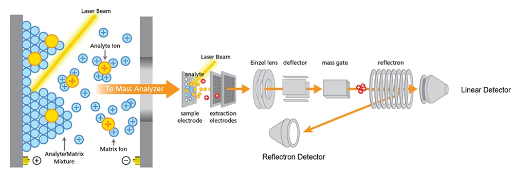
Figure 4. Path of ions after ionization through a time-of-flight sequence to a linear or reflectron detector.
III. TOF/TOF CID Analysis
Peptide fragmentation is a tool that can be used to determine the sequence of a peptide or understand specific locations of amino acid modifications. There are three types of fragmentation in MALDI analysis, In-source decay (ISD), post source decay (PSD) and collision induced dissociation (CID). For these methods, the fragmentation of the analyte occurs at different points of the extraction cycle. ISD produces ion fragments of the analyte before or when they are extracted from the ion source. This can be due to a high laser power or the ease of fragmentation of a specific analyte. More controlled fragmentation can be achieved with PSD and CID. For these techniques, the ion of interest can be isolated using an ion gate and then subjected to further fragmentation. PSD is characterized by the fragmentation of ions in the flight tube after they have been extracted from the source. While in CID, ions are accelerated in a collision cell where they can collide with a neutral gas to produce fragments.
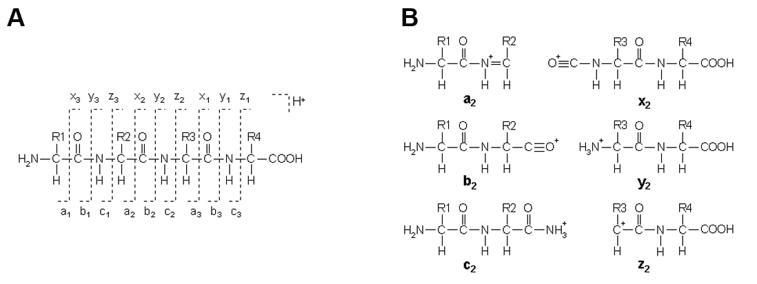
Figure 5. (A) Generic peptide showing the positioning for the formation of a, b, or c ions with charges on the N terminal fragment, and x, y, or z ions where charges fall on the C terminal fragment. (B) Resulting a, b, and c ions, holding charges on the N terminus and x, y, and z ions with charges on the C terminus. Structures include a single charge carrying proton. In other forms of ionization, peptides carry two or more charges, resulting in fragment ions with more than one proton.
Once a precursor ion has been selected and isolated by the ion gate, it enters the collision cell where it collides with neutral gas molecules. These collisions cause an energy transfer to the precursor ion and lead to fragmentation, i.e. fragment ions. The product ions enter a second source where they are reaccelerated and refocused before being detected. Collision induced fragmentation of peptides will generate product ions that result from the cleavage of the amino acid backbone or side chain. The most common cleavage occurs on the amide bond (CO-NH). The nomenclature of the product ion considers the localization of the positive charge after the cleavage. If the positive charge remains on the N-terminal side, the product ion is called a b ion. If the charge is located on the C-terminal end, the product ion is known as a y ion (figure 5). Interpretation of the resulting peptide fragments has been simplified using different programs that calculate the expecting mass of the b and y ions that could be produced for each sequence.
IV. MALDI mini – Digital Ion Trap
(Iwamoto, S., Hosoi, K., Furuta, M. Shichi, H., Yamauchi, S. Nishikaze, T., Nakaya, S., Watanabe, K., Kodera, K., Tanaka, K., Development of MALDImini-1 Digital Ion Trap Mass Spectrometer, 2020, 77:1 ,2-12.)
The MALDImini differs from traditional MALDI-TOF analyzers for two main reasons, first being the use of a digital ion trap (DIT) and second being the lack of a flight tube. The DIT technology provides an alternate mode of ion gating and ion trapping, along with the ability to handle MS/MS and MS3 analysis without the need for an added collision cell. The lack of a flight tube greatly reduces the size of the instrument, in combination with a smaller optic system and the DIT, the MALDImini has a footprint equivalent to an A3 sheet of paper. The analysis principals are the same ones MALDI-TOF uses. The analyte and matrix mixture excited with a laser source to generate a gas plume containing analyte ions of interest. The gas plume is directed towards a quadrupole ion deflector that redirects ions towards the ion trap and ultimately the detector.
The DIT was developed by Shimadzu Corporation. The ion trap is driven by rectangular wave electric fields instead of a sinusoidal electric field used for a conventional radio frequency (RF) ion trap. The rectangular waves resemble a digital signal, causing it to be called a digital ion trap (figure 6). The DIT in the MALDImini is a three-dimensional quadrupole ion trap (3D-QIT), and is configured as a ring electrode between a pair of endcap electrodes (figure 7). The use of a DIT allows for ion trapping and ejection for MS/MS and MS3 analysis. This is through a digital asymmetric wave isolation (DAWI), a technique unique to DIT technology. Through a change in the rectangular voltage, the mass scan line is sloped so that only ions in the specified mass range remain in the trap space while all others are ejected. This makes it incredibly useful for MS/MS and MS3 analysis as specific ions can be selected for further analysis. Fragmentation of selected ions occurs by collision with argon. The selected ions have increased kinetic energy and induces ion dissociation by collision with the gas.

Figure 6. A conventional radiofrequency with a sinusoidal wave that requires an increased voltage to modulate scan. A DIT waveform uses rectangular waves, allowing for a lower energy source to increase the frequency of the resulting waveform.
The MALDImini has three collection modes, MS1, MS2 and MS3. The MS1 mode is a typical MALDI-TOF spectra, displaying the mass of the parent ion. MS2 allows users to select a parent ion found in the MS1 spectra to fragment with the help of the argon collision gas. Further fragmentation can be observed in MS3, where a peak detected in MS2 can be subjected to further fragmentation. MSn modes have been used for structural analysis of modified peptides, where a modification such as phosphorylation or glycosylation can be interrogated in MS2. The resulting unfunctionalized peptide peaks can be further interrogated in MS3 as a mode for sequence determination. This is just an example of one of many possible uses of the MSn modes on the MALDImini.
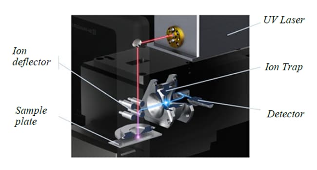
Figure 7. Internal component configuration for the MALDImini-1.
V. Atmospheric MALDI
MALDI can occur under atmospheric conditions and under vacuum. Both forms of ionization come with their own advantages and disadvantages with respect to analyte classes and matrix stability. Atmospheric Pressure MALDI (AP-MALDI) features the generation of an ion plume under atmospheric conditions before a push voltage directs ions toward a vacuum-controlled environment for mass detection.18 In vacuum MALDI, ions are formed on a flat charged surface with an extraction region that accelerates the ions towards the drift region.19 The formation of ions on a flat surface allows for the elimination of spatial spread, helping to create a more uniform spectrum. Early analysis showed that AP-MALDI sources were better at removing matrix adducts compared to their vacuum MALDI adducts.19 AP-MALDI is a valuable tool for the detection of ions that are thermally labile or easily lost under vacuum ionization.20 In AP-MALDI, ion generation and movement towards the flight tube can occur when ions are desorbed into a stream of nitrogen gas and directed towards the mass spectrometer. This process is similar to the ion movement process of electrospray ionization (ESI). The sensitivity of AP-MALDI is dependent on the components that facilitate the transition of ions towards the mass analyzer.20 These include the geometry of the target tip, nitrogen gas flow rate, and position of the gas nozzle, to name a few.20 Additionally, source parameters and experimental conditions can affect analysis sensitivity. Early AP-MALDI analyzers utilized time-of-flight (TOF)18 and electrospray ion trap mass spectrometers.20, 21 The use of AP-MALDI opens new avenues for analyte detection that could otherwise be very challenging in vacuum. 21, 22 A diagram of an AP-MALDI instrument, the IMScope QT is shown in figure 8.
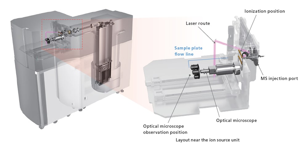
Figure 8. Diagram of the AP-MALDI configuration in the IMScope QT. Sample ionization occurs near the MS injection port to facilitate transfer of ions to LCMS-9030 mass detector.
AP-MALDI has been used to analyze a variety of compounds including tryptic peptides23, 24, 25, oligosaccharides26 and proteolytic fragments.27 AP-MALDI has also been a valuable tool for MALDI based mass spectrometry imaging (MSI) experiments28, offering higher spatial resolution29 compared to some vacuum MALDI-MSI.30 Using AP-MALDI for imaging experiments allows the use of different MALDI matrices that would otherwise sublimate under a high vacuum environment, enabling the detection of volatiles and other analytes that could be challenging to see vacuum stable matrices.31 A promising MALDI matrix for detection of lipids, 2,6-dihydroxyacetophenone or 4-nitroaniline is not stable under vacuum imaging experiments. These compounds sublimate under high vacuum conditions, leading to a continuous change in matrix/analyte ratios during imaging experiments.32 While there are advantages and disadvantages to using AP-MALDI over vacuum MALDI for different analysis, the addition of AP-MALDI is a valuable and complementary tool in the ever-evolving research space.
[1] Karas, M.; Bachman, D.; Bahr, U.; Hillenkamp, F. Matrix-assisted ultraviolet laser desorption of non-volatile compounds, Int. J. Mass Spectrom. Ion Processes 1987, 78, 53-68
[2] Karas, M.; Hillenkamp. F. Laser Desorption Ionization of Proteins with Molecular Masses Exceeding 10,000 Daltons. Analytical Chemistry. 1988, 60, 2299-2301
[3] Tanaka, K,; Waki, H; Ido, Y.; akita, S.; Yoshida, Y.; Yoshida, T. Protein and polymer analyses up to m/z 100 000 by laser ionization time-of-flight mass spectrometry, Rapid Commun. Mass Spectrom. 1988, 2, 151-153
[4] Dreisewerd, K. The desorption process in MALDI. Chemical Reviews. 2003, 103, 395-426
[5] Knochenmuss, R.; Zenobi, R. MALDI ionization: the role of in-plume processes. Cehmical Reviews 2003, 103, 441-452
[6] Karas, M.; Kruger, R. Ion formation in MALDI: The cluster ionization mechanism. Chemical Reviews 2003, 103, 441-452.
[7] Zhang, W.; Niu, S.; Chait, B. Exploring Infrared Wavelength Matrix-Assisted Laser Desorption/Ionization of
Proteins with Delayed-Extraction Time-of-Flight Mass Spectrometry, J Am Soc Mass Spectrom 1998, 9, 879-884
[8] Jaskolla, T.; Karas, M. Compelling Evidence for Lucky Survivor and Gas Phase Protonation : The Unified MALDI Analyte Protonation Mechanism, J Am Mass Spectrom 2011, 22, 976-988.
[9] Knochenmuss. R. MALDI Ionization Mechanisms: an Overview. From electrospray and MALDI mass spectrometry: Fundamentals, instrumentation, Practicalities and Biological Applications, Second edition, Edited by Richard B. Cole. 2010, pages 149-175. (c) 2010 John Wiley & Sons, Inc.
[10] Cotter, R. MALDI Ionization Mechanisms: an Overview. From electrospray and MALDI mass spectrometry: Fundamentals, instrumentation, Practicalities and Biological Applications, Second edition, Edited by Richard B. Cole. pages 345-364. (c) 2010 John Wiley & Sons
[1]1 Jaskolla, T.; Karas, M. Compelling Evidence for Lucky Survivor and Gas Phase Protonation : The Unified MALDI Analyte Protonation Mechanism, J Am Mass Spectrom 2011, 22, 976-988.
[12] Price, D. Time-of-flight Mass Spectrometry Time-of-Flight Mass Spectrometry 1994, Chapter 1, 1-15.
[13] Kovtoun, S.; Cotter, R. Mass-Correlated Pulsed Extraction: Theoretical Analysis and Implementation With a Linear Matrix-Assisted Laser Desorption/Ionization Time of Flight Mass Spectrometer. J Am Soc Mass Spectrom, 2000, 11, 841-853.
[14] Cotter, R. MALDI Ionization Mechanisms: an Overview. From electrospray and MALDI mass spectrometry: Fundamentals, instrumentation, Practicalities and Biological Applications, Second edition, Edited by Richard B. Cole. pages 345-364. (c) 2010 John Wiley & Sons
[15] Mamyrin, B. A.; Karataev, V. I.; Shmikk, D. V.; Zagulin, V. A. Mass reflection: a new nonmagnetic time-of-flight high resolution mass-spectrometer, Soviet Physics JETP 1973, 37, 45
[16] Cornish, T., Bryden, W. Miniature Time-of-Flight Mass Spectrometer for a Field-Portable Biodetection System.Johns Hopkins APL Technical Digest, 1999, 20:3, 335-342.
[17] Matrix Science Peptide fragmentation. http://www.matrixscience.com/help/fragmentation_help.html (accessed 2023-03-03)
[18] Moyer, S.C.; Cotter, R.J. Atmospheric Pressure MALDI. Anal. Chem. 2002
[19] Cotter, R. MALDI Ionization Mechanisms: an Overview. From electrospray and MALDI mass spectrometry: Fundamentals, instrumentation, Practicalities and Biological Applications, Second edition, Edited by Richard B. Cole. Page 208. (c) 2010 John Wiley & Sons
[20] Laiko, V.V.; Moyer, S.C.; Cotter, R.J. Atmospheric Pressure MALDI/Ion Trap Mass Spectrometry Anal. Chem. 2000, 72, 5239-5243.
[21] Laiko, V.V.; Taranenko, N.I.; Berkout, V.D.; Musselman, B.D.; Doroshenko, V.M. Atmospheric pressure laser desorption/ionization on porous silicon. Rapid Commun. Mass Spectrom. 2002, 16: 1724-1742.
[22] Keller, C.; Maeda, J.; Jayaraman, D.; Chakraborty, S.; Sussman, M.R.; Harris, J.M.; Ane, J-M.; Li, L. Comparison of Vacuum MALDI and AP-MALDI Platforms for the Mass Spectrometry Imaging of Metabolites Involved in Salt Stress in Medicago truncatula. Front. Plant Sci. 2018, 9.
[23] Schneider BB, Lock C, Covey TR. AP and vacuum MALDI on a QqLIT instrument. J Am Soc Mass Spectrom. 2005 Feb;16(2):176-82.
[24] Gudlavalleti SK, Sundaram AK, Razumovski J, Doroshenko V. Application of atmospheric pressure matrix-assisted laser desorption/ionization mass spectrometry for rapid identification of Neisseria species. J Biomol Tech. 2008 Jul;19(3):200-4.
[25] Nguyen J, Russell SC. Targeted proteomics approach to species-level identification of Bacillus thuringiensis spores by AP-MALDI-MS. J Am Soc Mass Spectrom. 2010 Jun;21(6):993-1001.
[26] Chong SL, Nissilä T, Ketola RA, Koutaniemi S, Derba-Maceluch M, Mellerowicz EJ, Tenkanen M, Tuomainen P. Feasibility of using atmospheric pressure matrix-assisted laser desorption/ionization with ion trap mass spectrometry in the analysis of acetylated xylo oligosaccharides derived from hardwoods and Arabidopsis thaliana. Anal Bioanal Chem. 2011 Nov;401(9):2995-3009.
[27] Grasso G, Rizzarelli E, Spoto G. AP/MALDI-MS complete characterization of the proteolytic fragments produced by the interaction of insulin degrading enzyme with bovine insulin. J Mass Spectrom. 2007 Dec;42(12):1590-8.
[28] Dilmetz, BA, Lee, Y-R, Condina, MR, et al. Novel technical developments in mass spectrometry imaging in 2020: a mini review. Anal Sci Adv. 2021; 2: 225–237.
[29] Kompauer M, Heiles S, Spengler B. Atmospheric pressure MALDI mass spectrometry imaging of tissues and cells at 1.4-μm lateral resolution. Nat Methods.2017;14(1):90-96.
[30] Barry J A, Ait-Belkacem R, Hardesty W M ,et al. Multicenter validation study of quantitative imaging mass spectrometry. Anal Chem.2019;91(9):6266-6274
[31] Islam, A.; Sakamoto, T.; Zhai, Q.; Rahman, M.M.; Mamun, M.A.; Takahashi, Y.; Kahyo, T.; Setou, M. Application of AP-MALDI Imaging Mass Microscope for the Rapid Mapping of Imipramine, Chloroquine, and Their Metabolites in the Kidney and Brain of Wild-Type Mice. Pharmaceuticals 2022, 15, 1314.
[32] Leopold J, Popkova Y, Engel KM, Schiller J. Recent Developments of Useful MALDI Matrices for the Mass Spectrometric Characterization of Lipids. Biomolecules. 2018 Dec 13;8(4):173.


